Tunneling Nanotubes in Motion
Movie 1. Movement of GFP-Myo10 within TNTs
CADs cells transiently transfected with GFP-Myo10 for 24 hrs were imaged in a temperature and CO2 controlled chamber with a microscope confocal Revolution Nipkow spinning-disk imaging system (Andor Technology). Every 30 sec, multiple Z-stacks were acquired and projections were obtained for each time point using the ImageJ software. Over time, GFP-Myo10 can be seen moving back and forth within TNTs. Gousset et al., J Cell Sci. 2013 Jul 25.
Movie 2. Cell-to-cell detachment mechanism of TNT formation in CAD cells
Unlabeled CAD cells were put into the incubation chamber of a Biostation IM from Nikon for time-lapse analysis. Phase contrast images were taken every minute for 12 hours. Over time, multiple TNTs resulting from cell-to-cell detachments can be observed. Gousset et al., J Cell Sci. 2013 Jul 25.
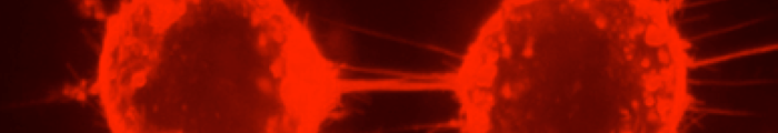
Movie 3. Filopodia-driven mechanism of TNT formation in CAD cells
CAD cells were labeled with 5-carboxytetramethylrhodamine succinimidyl-ester (for membrane labeling) for 1h at 37°C and imaged over time, every 30 sec for 40 minutes with a microscope confocal Revolution Nipkow spinning-disk imaging system (Andor Technology). For each time point, multiple Z-stacks were acquired. Because of the movement of the cells, Z-stacks required readjustments over time and this movie was made from concatenated movies. Projections were obtained for each time point using the ImageJ software. Over time, the dorsal filopodia can be seen moving toward the acceptor cells. After TNT attachment, the structure remained stable for hours. Gousset et al., J Cell Sci. 2013 Jul 25.